|
Discover your next important protein interaction
with this new Pierce Far-Western Kit
ProFound Far-Western Biotinylated-Protein:Protein
Interaction Kit
The first nonisotopic far-Western Protein:Protein
Interaction Kits that detect interactions in-gel or
on-membrane.
Far-Western protein:protein interaction analysis need
not be difficult or mysterious. The new ProFound
Far-Western Biotinylated-Protein:Protein Interaction Kit
represents a novel nonisotopic approach to far-Western
analysis that can be performed either directly in the
gel or, like the classical method, on a membrane using
sensitive chemiluminescent detection.
Far-Western Primer
A far-Western is a method designed to detect protein
interactions. In a classical far-Western strategy,
proteins are first transferred from gel to membrane.
These proteins are then probed with a pure protein known
to interact or putatively interact with one or more
proteins transferred to the membrane. The protein probe
or "bait" protein is isotopically labeled and the
interaction with the "prey" protein is detected on the
membrane. Since the probe in far-Western analysis is
not an antibody, as it would be in classical
Western blot detection, the term "far" was
adopted to make this distinction. Therefore, the term
far-Western emerged to describe a very useful
in vitro protein:protein interaction method.
ProFound Far-Western Protein:Protein Interaction
Kit
A Nonisotopic Approach to Far-Western Analysis
In-Gel or On-Membrane
|
ProFound Far-Western
Biotinylated-Protein:Protein Interaction
Kit |
|
This kit utilizes
biotin modification of a purified "bait" protein
probe. Prey proteins separated in-gel or
transferred to a membrane can be probed with this
biotinylated bait. Detection of an interaction
with "prey" protein(s) is achieved with a
streptavidin-horseradish peroxidase conjugate and
a chemiluminescent
substrate. |
How does this far-Western kit work?
Exclusive Pierce UnBlot Technology
Adopted for Detection of Protein
Interactions
The far-Western protein:protein interaction kit
incorporates the major advantages inherent in the Pierce
UnBlot In-Gel Chemiluminescent Detection Platform,
including the subsequent elimination of the need for
protein transfer. Interactions can be directed directly
in-gel with the appropriate probe. In addition,
methodology is provided that enables use of the kit for
on-membrane applications if additional sensitivity is
needed to detect the interaction of interest.
Optimization may be required to obtain the best results
in a given system.
Kit Applications
· Protein:Protein
Interaction Discovery
Direct in-gel or on-membrane detection of a
potential interaction using a purified
biotinylated-protein as a probe
Compared to classical far-Western detection on a
membrane, the in-gel technology incorporated into this
kit is a faster and easier method to reveal potential
interactions.
In those instances where an interaction cannot be
readily detected in-gel, the reagents provided with this
kit can also be used for on-membrane
(nitrocellulose/PVDF) detection.
The strategy employed aids in the detection of strong
to moderate interacting pairs. Weak or transient
interactions may not be readily observed with these
probes either in-gel or on-membrane.
· Protein:Protein
Interaction Confirmation
The far-Western based kit can be used as an in
vitro tool for the verification or confirmation of
known or putative bait:prey interacting pairs.
Far-Western Biotinylated-Protein:Protein
Interaction Kit Results
Confirmation of a Known Protein Interactions with
a Biotinylated Bait.
In-gel detection using UnBlot Methodology vs.
classical on-membrane detection
|
1 2 3 |
1 2 3 |
1 2 3 |
1 2 3 |
|
|
|
|
|
|
1A)
In-gel
detection |
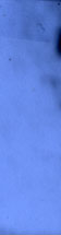
1B)
In-Gel
Control |
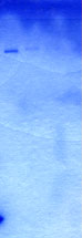
1C) Total
Protein
Stain |
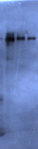
1D)
Membrane
detection |
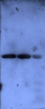
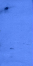
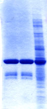
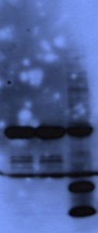
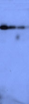
Figure 1. Protein:Protein Interaction – Far
Western In-Gel and Membrane. Purified BIR2 and BIR2
bacterial lysate samples were separated by SDS-PAGE on
4-20% Tris-Glycine gels. Two gels (1A and 1B) were
probed and processed using in-gel far-Western detection
and UnBlot Technology-based method. A third gel was
transferred to a nitrocellulose membrane and processed
using the classical far-Western membrane-based probing
and detection. Gel 1A)
The samples were probed in-gel with purified
Biotinylated-Smac diluted 1:100 followed by
Streptavidin-HRP diluted 1:20,000.
Gel 1B) The control
gel was probed with non-biotinylated purified Smac
followed by Streptavidin-HRP. Gel 1C) Following the in-gel far-Western
detection, one gel was stained for total protein with
GelCode Blue Stain Reagent (Product # 24590). Gel 1D) The membrane was probed
with purified Biotinylated-Smac diluted 1:200 followed
by Streptavidin-HRP diluted 1:250,000.
Pierce UnBlot Chemiluminescent Substrate Working
Reagent was used for the detection of 1A, 1B and 1D
shown above. Lane 1 and 2 correspond to purified BIR2,
1.7 µg and 3.1 µg, respectively. Lane 3 corresponds to
BIR2 bacterial lysate. The biotinylation of Smac was
done using Sulfo-NHS-LC-Biotin (Product # 21335). BIR2
(GST-BIR2)- Binding Domain of XIAP Smac (9xHis-Smac)-
Second Mitochondrial-Derived Activator of Caspases
|
1 2 3 |
1 2 3 |
1 2 3 |
1 2 3 |
|
|
|
|
|
|
2A. |
2B. |
2C. |
2D. |
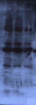
Figure 2. Chaperone Proteins: Interaction
of p90 with Biotinylated-Hop. Chaperone protein p90
(denatured) was separated by SDS-PAGE on a 10-20%
Tris-Glycine gel. Lane 1, 2 and 3 correspond to 15 µg,
30 ng and 15 ng, respectively, of p90 chaperone protein.
Gel 2A) In-gel far-Western detection. Gel
2A was probed with Biotinylated-Hop followed by
Streptavidin-HRP. Gel 2B) In-gel far-Western
detection control. Gel 2B was probed with
non-biotinylated Hop followed by Streptavidin-HRP.
Gel 2C) Total protein staining. Following the
in-gel immunodetection procedure in 2A and 2B above, one
gel was stained with GelCode Blue Stain Reagent, a
Coomassie Dye-based reagent. Gel 2D)
Membrane far-Western detection. A third gel was run and
transferred to a nitrocellulose membrane. The membrane
was probed with Biotinylated-Hop followed by
Streptavidin-HRP. The protein was detected with UnBlot
Substrate Working Reagent. The biotinylation of Hop was
done using Sulfo-NHS-LC-Biotin (Product # 21335).
|
1 2 3 |
1 2 3 |
|
|
|
|
3A. |
3B. |
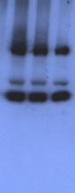
Figure 3. Probing for interactions in-gel
with a biotinylated whole cell lysate.
Mycobacterium avium complex (MAC101) lysate
samples were separated by SDS-PAGE on two 8-16%
Tris-Glycine gels (3A and 3B). Lanes 1, 2 and 3
correspond to 5 µg, 30 µg and 15 µg of total MAC101
lysate protein, respectively. Gel 3A) Gel was
incubated with a 1:100 dilution of Biotinylated-Human
epithelial cell lysate (B-Hep2 cell lysate). Starting
protein concentration was estimated at 2 mg/ml.
Gel 3B) Negative control gel was incubated
in the appropriate dilution buffer alone. Gels 3A and 3B
were developed using Streptavidin-HRP (at a 1:25,000
dilution of a 1 mg/ml solution) and UnBlot Substrate
Working Reagent.
Features
|
- In-gel or
on-membrane detection
options
|
- Kit can be adapted
to in-gel or on-membrane detection. The in-gel
detection method offers speed vs. the higher
sensitivity of on-membrane
detection
|
|
- Nonradioactive
alternative for far-Western
analysis
|
- Reliable and
sensitive biotin:streptavidin-HRP chemistry
combined with chemiluminescent detection offer a
practical and safe alternative to radio-labeling
the bait protein
|
|
|
- Kit targets moderate
to strong associations between a prey and the
biotinylated bait protein
probe
|
|
- Primary
antibody-free detection
|
- Interactions can be
detected with systems that eliminate the need to
raise an antibody initially to the probe
protein
|
- In-gel method saves
time and cost
|
- No transfer or
blocking steps reduces detection time after
electrophoresis
- Costly transfer
units, transfer buffers, transfer membranes and
filter papers are not required if interactions
can be detected
in-gel
|
|
|
- In-gel detection
technology prevents problems associated with
incomplete or inefficient transfer making more
of the potential prey protein available to the
bait protein probe
|
|
- Compatible with both
SDS-PAGE and native gels
|
- Provides option to
probe for prey proteins in a more native
environment as reduced or denaturing systems may
not always present an interface that promotes
the intended interaction
|
|
- Uniform presentation
of prey protein
|
- In-gel detection
avoids problems associated with uneven transfer
[e.g., low molecular weight (M.W.) proteins
transfer more efficiently than higher M.W.
proteins]
|
|
- Reduced nonspecific
binding
|
- For example,
Biotin/Streptavidin–HRP systems demonstrate less
nonspecific binding compared to antibodies
directed against the bait
protein
|
|
- Compatible with
protein staining
|
- The gel can be used
for total protein staining; e.g., GelCode Blue
Stain Reagent after the chemiluminescent
detection step.
- No need to run 2
gels
|
|
- Sensitive to 5 ng
prey protein in-gel
|
- Comparable to ECL™
Substrate on a membrane
- The sensitivity may
vary by the interaction under study and the
matrix upon which it is being
detected
|
References
- Towbin, H., Staehlin, T. and Gordon, J. (1979).
Electrophoretic transfer of proteins from
polyacrylamide gels to nitrocellulose sheets:
Procedure and some applications. Proc. Natl. Acad.
Sci. USA 76, 4350-4354.
- Renart, J., Reiser, J. and Stark, G.R. (1979).
Transfer of proteins from gels to diazobenzyloxymethyl
paper and detection with antisera. A method for
studying antibody specificity and antigen structure.
Proc. Natl. Acad. Sci. USA 76, 3116-3120.
- Burgess, R., Arthur, T.M. and Pietz, B.C. (2000).
Mapping protein-protein interaction domains using
ordered fragment ladder far-Western analysis of
hexahistidine-tagged fusion proteins. Meth. Enzymol.
328, 141-157
- Lin, W. and Kasamatsu, H. (1983). On
electrotransfer of polypeptides from gels to
nitrocellulose membranes. Anal. Biochem. 128, 302-311.
- DenHollander, N. and Befus, D. (1989). Loss of
antigens from immunoblotting membranes. J. Immunol.
Meth. 122, 129-135.
- Reddy, V.M. and Kumar, B. (2000). Interactions of
Mycobacterium avium complex with human respiratory
epithelial cells. J.I.D 181,1189-1193.
Ordering Information
|
 Instruction Book Instruction Book  MSDS MSDS |
| Buy |
Product # |
Description |
Pkg. Size |
Files |
Price |
| Add |
23500 |
ProFound Far-Western
Biotinylated-Protein:Protein Interaction
Kit
Streptavidin-Horseradish Peroxidase
(Streptavidin-HRP), 0.1 mg,
lyophilized
Dilution Buffer (10X), 50
ml
BupH Phosphate Buffered Saline Packs, 17
packs, 0.1 M phosphate, 0.15 M NaCl, pH
7.2
UnBlot Luminol Enhancer, 55 ml
UnBlot
Stable Peroxide, 55 ml
Surfact-Amps 20, 6 x
10 ml vials, 10% solution
Pre-Cut Cellophane,
10 sheets |
kit |
|
$335.00 | |
|
|